歡迎來到匠仁醫療設備有限公司網站!


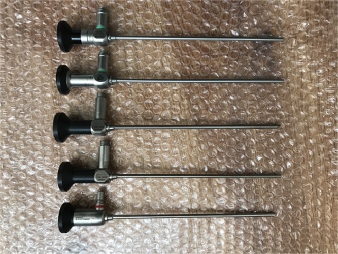
內窺鏡是1853年法國醫生德索米奧創制的。內窺鏡是一種常用的醫療器械。由頭端、彎曲部、插入部、操作部、導光部組成。使用時先將內窺鏡導光部接到配套的冷光源上,然后將插入部導入預檢查的器官,控制操作部可直接窺視有關部位的病變。
The first endoscope in the world was created by French doctor Desomio in 1853. Endoscope is a commonly used medical device. Composed of head end, bending part, insertion part, operation part, and light guide part. When using, first connect the endoscope's light guide to the matching cold light source, and then guide the insertion part into the pre examination organ to control the operation department to directly observe the lesions in the relevant areas.

早的內窺鏡被應用于直腸檢查。醫生在病人的肛門內插入一根硬管,借助于蠟燭的光亮,觀察直腸的病變。這種方法所能獲得的診斷資料有限,病人不但很痛苦,而且由于器械很硬,造成穿孔的危險很大。盡管有這些缺點,內窺鏡檢查一直在繼續應用與發展,并逐漸設計出很多不同用途與不同類型的器械。
The earliest endoscopes were used for rectal examination. The doctor inserted a hard tube into the patient's anus and observed the lesion in the rectum with the light of a candle. The diagnostic data obtained by this method is limited, and patients not only suffer greatly, but also have a high risk of perforation due to the hard instruments. Despite these drawbacks, endoscopic examination has continued to be applied and developed, gradually designing many different types of instruments for different purposes.
1855年,西班牙人卡赫薩發明了喉鏡。德國人海曼·馮·海莫茲于1861年發明了眼底鏡。
In 1855, the Spaniard Cahesa invented the laryngoscope. German Heiman von Helmholtz invented the ophthalmoscope in 1861.
1878年,愛迪明了燈泡,特別是出現微型燈泡后,使內窺鏡有了很大發展,臨時安排的手術內窺也可達到非常精確的程度。
In 1878, Edison invented the light bulb, especially with the emergence of miniature light bulbs, which greatly developed endoscopes. Temporary surgical endoscopes can also achieve a very precise level.
1878年德國泌尿科姆·尼茲創造了膀胱鏡,用它可以檢查膀胱內的某些病變。
In 1878, German urologist M Niz created cystoscopy, which can examine certain lesions in the bladder.
1897年,德國人哥·基利安設想支氣管鏡。
In 1897, a German named G ? rgen envisioned bronchoscopy.
1862年,德國人斯莫爾創造了食道鏡。
In 1862, German Smoll created the esophagoscope.
1903年,美國人凱利創制了直腸鏡,但是到1930年后才開始普遍使用。
In 1903, American Kelly created the concept of colonoscopy, but it was not until 1930 that it became widely used.
1913年,瑞典人雅各布斯改革了胸膜鏡檢查法。
In 1913, Swedish scholar Jacobs reformed the pleural examination method.
1922年,美國人欣德勒創立了胃鏡檢查法。
In 1922, American Schindler founded the gastroscopy method.
1928年,德國人卡爾克創立了腹鏡檢查法。
In 1928, German Carl K established the abdominal endoscopy method.
1936年,美國人斯卡夫進行了腦室鏡檢試驗,直到1962年,才由德國人古奧和弗累斯梯爾創立了腦室鏡檢法。從此形成一整套鏡檢法系列。
In 1936, an American named Scarf conducted a ventriculoscopic examination, and it was not until 1962 that the method of ventriculoscopic examination was established by German authors G ü o and F ü rster. From then on, a complete series of mirror inspection methods was formed.
1963年,日本開始生產纖維內窺鏡,
In 1963, Japan began producing fiber endoscopes,
1964年研制成功纖維內窺鏡的活檢裝置,這種取活檢的特別活檢鉗能夠有合適的病理取材而且危險小。
In 1964, a biopsy device using a fiber endoscope was successfully developed. This special biopsy forceps for biopsy can provide suitable pathological samples with minimal risk.
1965年,纖維結腸鏡制成,擴大了對于下消化道疾病的檢查范圍。
In 1965, fiber colonoscopy was made, expanding the scope of examination for lower gastrointestinal diseases.
1967年開始研究放大纖維內窺鏡以觀察微細病變。光纖內窺鏡還可以用來做體內化驗,如測量體內溫度、壓力、移位、光譜吸收以及其他數據。
In 1967, research began on magnifying fiber endoscopes to observe microscopic lesions. Fiber optic endoscopes can also be used for in vivo laboratory tests, such as measuring internal temperature, pressure, displacement, spectral absorption, and other data.
1973年,激光技術應用于內窺鏡的上,并逐漸成為經內窺鏡有消化道出血的手段之一。
In 1973, laser technology was applied to the treatment of gastrointestinal bleeding through endoscopy and gradually became one of the methods for treating gastrointestinal bleeding through endoscopy.
1981年,內窺鏡超聲波技術研制成功,這種把的超聲波技術與內窺鏡結合在一起的新發展,大大增加了對病變診斷的準確性。
In 1981, endoscopic ultrasound technology was successfully developed, which combined advanced ultrasound technology with endoscopy and greatly increased the accuracy of lesion diagnosis.
 公司:匠仁醫療設備有限公司
 樊經理:13153199508 李經理:13873135765
公司地址:山東省濟南市槐蔭區美里東路3000號德邁國際信息產業園6號樓101-2室 湖南省長沙市雨花區勞動東路820號恒大綠洲14棟2409室